Design of Multifunctional Nanoparticles for Cellular Labeling and Tracking, Delivery and Scavenging of Active Substances in vivo
Gulin-Sarfraz, Tina (2019-05-14)
Gulin-Sarfraz, Tina
Åbo Akademi - Åbo Akademi University
14.05.2019
Publikationen är skyddad av upphovsrätten. Den får läsas och skrivas ut för personligt bruk. Användning i kommersiellt syfte är förbjuden.
Publikationens permanenta adress är
https://urn.fi/URN:ISBN:978-952-12-3819-2
https://urn.fi/URN:ISBN:978-952-12-3819-2
Abstrakt
Nanomedicine, the application of nanotechnology in the biomedical field, has gained tremendous interest for improving diagnostic and therapeutic interventions with potential in personalized medicine. The aim of personalized medicine is to guarantee that the most suitable medicine is given to each patient, and is further efficiently and safely delivered to the right place of treatment in the body. The research within the diverse field of nanomedicine is steering towards this goal; with high expectations of novel delivery systems for commercially available drugs, tailored for individual patients pre-selected by sophisticated diagnostic tools, and in addition, targeted to a specific site of action with subsequent controlled drug release. To potentially reach this goal, the preparation and modification steps of the nanomedicines have to step-wise be carefully considered, and further studied and evaluated for their specific applications in vivo.
In this thesis, the main emphasis was to design and construct multifunctional silica-based nanoparticles and evaluate them for specific diagnostic and therapeutic applications. The influence of different surface coatings was thoroughly investigated for the effect of stabilizing and dispersing the nanoparticles, and subsequently for maximizing cellular internalization. This effect was further explored by developing magnetite-silica core-shell particles and evaluating the synergistic effect of surface coating and an external magnetic field on the cellular uptake, in addition to the performance of the particles as contrast agents for magnetic resonance imaging (MRI).
Diagnostic imaging techniques, such as MRI, are becoming increasingly important for early diagnosis of various pathologies. To improve the quality of generated images, as well as to visualize and track cells (e.g. stem cells after implantation), novel imaging probes are required. Here, porous silica nanoparticles were evaluated to serve as “nano-containers” for carrying a high amount of commercially available fluorescent imaging agents, for long-term cellular tracking. A tumor of pre-labeled breast cancer cells was monitored in vivo for a period of one month, and real-time detection of circulating metastatic cells was demonstrated. The same properties that render porous silica nanoparticles good candidates to serve as carriers for imaging agents, are also applicable for drugs. This approach was utilized by loading a large amount of anti-inflammatory drug into the particles, and the therapeutic treatment effect on airway inflammation in mice was studied after pulmonary administration of the particles.
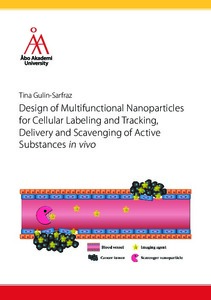
Molecular imaging probes intended for diagnostic imaging, are normally administered through intravenous injection and thus rapidly distributed all over the body. These contrast agents are typically small molecules capable of crossing the blood-tissue barriers and accumulating in tissues, especially when the barrier is disrupted as in the case of different pathological conditions. When imaging a pathological site, such as a tumor or inflammation, a problem is to accurately differentiate between the signals originating from the contrast agent in the blood circulation and the signals originating from surrounding tissue. Based on this, a nanoparticle-based scavenger-system for catching and quenching the signal of a circulating contrast agent (tracer) is demonstrated. The interaction between tracer and scavenger was investigated by photonic measurements, based on Förster/fluorescence resonance energy transfer (FRET). FRET is a useful tool to visualize short-distance molecular interactions. Thus, this approach is further utilized to develop a nanoparticle-based reporter system to study intracellular redox-induced delivery of an active model compound. Intracellular release can be particularly challenging to study, however highly desired, since many active compounds are non-toxic and can not be validated based on their cytotoxic action. A reportersystem may then serve as a tool for real-time monitoring of intracellular compound cleavage. The results presented in this thesis contribute in the process of developing tailored multifunctional nanoparticles for similar diagnostic and therapeutic applications as presented here.
Collections
- 317 Farmaci [18]
